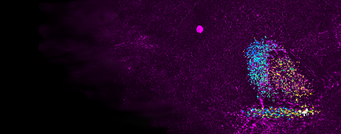

Expansion Microscopy Image Right
Left is a confocal image of normal Drosophila brain expressing a dopaminergic synaptic GFP marker (green) with a background stain (magenta). On the right is an expansion microscopy image taken under the same imaging conditions of a dopaminergic synaptic GFP (green) and membrane marker (red).